Posts

Showing posts with the label Medical Imaging

Doppler Ultrasound Is A Noninvasive Test For Estimating The Blood Flow Through Blood Vessels By Using Sound Waves

Image
  Doppler Ultrasound Doppler Ultrasound is a technology that has been developed for medical diagnostics since the 1950s. It has advanced to include real-time velocity measurements and flow mapping. It is now a common way to diagnose a variety of medical conditions. It is also a good option for assessing the severity of artery disease. Doppler Ultrasound is a test that uses sound waves to look at blood flow. It can show if there's blockage in a vein or artery, and it can help find blood clots or plaque buildup. It is noninvasive, usually lasts about an hour and has no side effects. A doctor might order a doppler ultrasound to diagnose problems with the blood vessels in abdomen, legs or arms. It can also be used to check for abnormal blood flow after surgery or treatment. During a Doppler Ultrasound , a technician presses a small handheld device called a transducer against the skin over the area of the body being scanned. The technician may apply a gel to the skin to make the ...

What is Positron Emission Tomography (PET) Scanners? Its uses and advantages?

Image
  Positron Emission Tomography (PET) Scanners A Positron Emission Tomography (PET) Scanners is an imaging test that uses radioactive substances to get images of internal organs. The tests help doctors diagnose a variety of medical conditions, including cancers and brain diseases. They are usually done along with CT or MRI scans to produce more detailed images. Positron Emission Tomography (PET) Scanners are different from X-rays, CTs, or MRIs in that they give information about blood flow and how body's cells use oxygen and sugar. This helps diagnose certain diseases and can tell doctors how a disease is progressing. A PET scan uses a radioactive tracer that is attached to sugar molecules. The tracer and the sugar molecules are injected into the body through an IV (intravenous) line. The tracers pass through the bloodstream and are picked up by areas in the body that have a higher metabolic rate. This helps the PET scanner create color-coded images of the body’s organs and tiss...

Intravascular Ultrasound Imaging System; Offer Valued Morphological Data For Cure And Treatment For CVDs

Image
  Intravascular Ultrasound Imaging System An Intravascular Ultrasound Imaging System is a tube-like diagnostic process that offers real-time visions of the structures inside and lining of the blood vessels. Doctors utilize this method to measure the amount of contraction or expansion of the artery and also to monitor atheroma that is concealed in the artery wall. A tube is inoculated into the artery and directed until it extents the region to be analyzed. The catheter is further linked to a small ultrasound transducer at an end and the other edge links to a computer terminal that changes sound waves released by the transducer into pictures on a monitor. Utilizing a huge-frequency ultrasound, Intravascular Ultrasound Imaging System allows the imagining of inner parts of blood vessels and atherosclerotic signs. Anyhow, it is restricted in its capability to detect the fibrous cap density and tissue combination of atherosclerotic abrasions. Additionally, the infiltration depth of h...

Intraoperative Imaging Technologies Are Rapidly Advancing With Continuous Technological Development To Support The Neurosurgical Tasks

Image
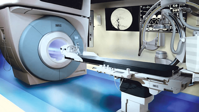
  Intraoperative Imaging Intraoperative Imaging is a powerful tool for neurosurgical guidance and can help minimize surgical risk and postoperative complications. However, implementing intraoperative imaging requires specialized training and expertise. A number of factors have to be considered, including patient risk and safety, cost, operator training, and additional time required for the procedure itself. Various imaging technologies are being developed to support the neurosurgical task. This includes real-time magnetic resonance imaging (MRI), intraoperative ultrasound, optical surface matching techniques, and other surgical navigation methods. Each of these has its own advantages and disadvantages, which should be taken into consideration when deciding on the most suitable approach to a specific surgical case. Incorporating Intraoperative Imaging into neurosurgery provides information about the location of the surgical site, the size and position of vessels and nerves, and...

Veterinary Imaging Is Advancing and It Is Widely Being Adopted With Increasing Awareness and Technological Developments

Image
  Veterinary Imaging Diagnostic imaging is a very important part of veterinary medicine. Veterinary Imaging allows veterinarians to identify and assess disease processes without invasive surgery or pain. It can also be a good way to get information about a pet's general condition. Radiography, ultrasound and computed tomography are a few of the most common types of diagnostic Veterinary Imaging in use today. The majority of the time, these procedures are used to examine a pet's abdominal and thoracic region. These scans provide a detailed anatomic picture of the organs and structures inside the animal's body. These images can help the veterinarian determine what is wrong with the animal, so they can better recommend treatment options. They are also an important tool for determining the stage of disease and helping with deciding whether or not surgery is needed. X-rays are the most popular form of radiography in veterinary medicine. This is due to they are fast and easy t...

Geriatric Population That Suffers From Disorders That Require Optical Imaging Devices, Which Will Contribute To The Growth Of The Optical Imaging Market

Image
  Optical imaging is a new non-invasive approach for obtaining images of the body's internal organs. Visible light and photons are used to provide a full image of interior organs and tissues, as well as images of tiny entities such as cells and molecules. The properties of photons, such as absorption and scattering, are used in optical imaging to measure and sense the qualities of organs and soft tissues. Despite the fact that optical imaging technology is still in its early stages of development, it offers a wide range of technological advantages over current imaging methods. Doctors use the images to diagnose and treat a variety of disorders, while scientists use them to conduct a variety of research projects.   According to the World Health Organization in 2012, the population over the age of 65 has risen the most and is anticipated to increase from 13% in 2010 to 53% by 2020. The high prevalence of neurological and ophthalmologic illnesses, increased R&D activities in ...

The Main Challenge In This Hyperspectral Imaging Market Is The High Cost Of These Systems, A Lack Of Awareness About Their Applications, A Scarcity Of Skilled Operators

Image
  Systems for hyperspectral imaging (imaging spectroscopy) are the result of extensive research and development efforts. These systems have a wide range of applications in industries such as healthcare, research and development, the food industry, the defence sector, and others such as remote sensing, calorimetry, and night vision. In terms of image quality, hyperspectral imaging differs from multispectral imaging in that it has a higher sensitivity than conventional or multispectral imaging. Multispectral imaging, for example, provides 2 to 20 images per dataset, whereas hyperspectral imaging provides more than 20 images per dataset. Furthermore, multispectral imaging provides 4 to 20 data points per pixel, whereas hyperspectral imaging provides the entire spectrum per pixel.   The leading factors driving the growth of the Hyperspectral Imaging Market are innovative products with advanced technology, high patient and operator safety, superior accuracy and consistency. The in...

In 2019, Imaging Systems Accounted For A Sizable Portion Of The Optical Imaging Market

Image
  Optical imaging is a new non-invasive technique for capturing images of the body's internal organs. Visible light and photons are used to create a complete image of internal organs and tissues, as well as images of smaller structures such as cells and molecules. The properties of photons, such as absorption and scattering, are used in optical imaging to measure and perceive the properties of organs and soft tissues. Despite the fact that optical imaging technology is still in its early stages of development, it offers a wide range of technological advantages over current imaging technologies. Doctors use the images to diagnose and treat a variety of diseases, and scientists use them to conduct a variety of research projects.   Because many cases are asymptomatic and do not undergo testing, the true number of infected people would be much higher than the official total. It is possible that the size of the optical imaging market has changed dramatically. In ophthalmology pract...

Growing Public Awareness Of Early Disease Diagnosis Will Be A Major Driving Factor In The Growth Of The Magnetic Resonance Imaging Systems (MRI) Market

Image
  Magnetic Resonance Imaging (MRI) is a sophisticated medical imaging technique used to image an individual's anatomy and physiological processes. MRI scanners create these images by employing high-power magnets, strong magnetic fields, radio waves, and magnetic field gradients. Over the forecast period, the increasing prevalence of chronic disorders is expected to drive growth in the global magnetic resonance imaging (MRI) market. According to the American Cancer Society, there will be an estimated 1,762,450 new cancer cases diagnosed and 606,880 cancer deaths in the United States in 2019. Furthermore, the growing geriatric population is expected to support the growth of the global magnetic resonance imaging (MRI) market.   The Magnetic Resonance Imaging Systems (MRI) Market is expected to benefit from the Asia Pacific region, where demand for magnetic resonance imaging systems is expected to be high due to a large proportion of the geriatric population residing in these cou...

Magnetic Resonance Imaging (MRI) System is a non-invasive imaging technique that creates comprehensive three-dimensional anatomy images.

Image
  Magnetic Resonance Imaging (MRI) is a sophisticated medical imaging technique used to visualise an individual's anatomy and physiological processes. MRI scanners use high-powered magnets, intense magnetic fields, radio waves, and magnetic field gradients to create these images. The launch of software to assist radiologists in MRI-guided operations is likely to provide lucrative growth prospects for worldwide magnetic resonance imaging (MRI) market competitors. For example, Qlarity Imaging, a producer of computer-aided breast cancer detection systems, stated in November 2019 that it would release supplementary software to aid radiologists in MRI-guided operations.  Magnetic Resonance Imaging (MRI) System is a non-invasive imaging technique that creates comprehensive three-dimensional anatomy images. It's frequently used to detect diseases, diagnose them, and track their progress. Magnetic resonance imaging (MRI) is a form of scan that produces detailed images of the inside o...

Optical Imaging solutions are used to obtain images of various body parts by virtue of photon characteristics

Image
Optical Imaging is an exciting new medical imaging technology. It is rapidly replacing traditional radiography as a procedure for revealing the internal structure of the body. Medical applications include the detection and diagnosis of disease using optical images taken using optical fibers. Other applications include mammography to detect breast cancer, ultrasound to detect tumors, and magnetic resonance imaging (MRI) to map the internal organs. Other uses include non-surgical imaging to detect congenital abnormalities in unborn children and evaluation of the abdomen, pelvic, and back regions to detect ovarian cancer. Imaging techniques are also used to reveal abnormal breasts in women with breast cancer, and to prevent scarring in patients who have undergone abdominoplasty. Medical imaging provides diagnostic and therapeutic benefits for a wide range of diseases and disorders. Traditional CT imaging is performed by injecting dye into the targeted areas. The dye penetrates the target...

What is Hyperspectral Imaging & Its Uses?

Image
  Systems for hyperspectral imaging (imaging spectroscopy) are the result of years of study and development. These systems are used in a variety of industries, including healthcare, research and development, food production, defence, and remote sensing, calorimetry, and night vision. Hyperspectral imaging differs from multispectral imaging in terms of image quality and sensitivity compared to conventional or multispectral imaging. Multispectral imaging, for example, produces 2 to 20 images per dataset, whereas hyperspectral imaging produces more than 20 images each dataset. Hyperspectral Imaging is a new spectroscopy-based analysis tool. For the same spatial area, it gathers hundreds of photos at various wavelengths. The data is organised into a hyperspectral cube, with two dimensions representing the scene's spatial extent and the third representing its spectral content. Hyperspectral images can be obtained from many different electromagnetic measurements. The most popular are vi...